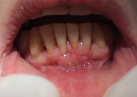
What is periodontitis (periodontitis)?
Periodontitis is a disease that affects all parts of the parodontium (tooth support device), the tissues in which the teeth are attached and which firmens the teeth in the jaw.
The tooth support device consists of:
- Gingiva – right
- Alveolar bone – a bone in which the tooth is located
- Periodontal fibers that connect the bone to the bone
- Cement root of the teeth.
The disease is destructive in nature and leads to a gradual, but progressive illness, and a final loss of one or more teeth. Studies have shown that about 50% of teeth have been extracted for periodontal disease, not for caries.
What are the clinical symptoms of periodontal disease?
Clinical symptoms occurring during periodontal disease are as follows:
- Inflammation of the gingival, ie inflammation of the right – bleeding gums
- pulling the right
- periodontal pockets
- purulent contents in the padorontal pockets
- loosening of teeth or betting
- pathological tooth migration – change of the position of the teeth, movement of the tooth to another place.
What is a periodontal pocket?
Periodontal pocket – PDP is the space between the gums and teeth where the deposits are filled with bacteria (connect the dental plaque with more extensive text) that act with their toxins, leading to inflammation of the soft wall of the pocket, ie, right and destruction of the bone cup in which the tooth is inserted. There is no pain during parodontopathy, and the symptoms are minor, except when complications occur.
The first signs of periodontitis – periodontitis
The first signs of periodontitis – periodontitis are subjective symptoms that the patient feels:
- bleeding from the right – which first occurs only for provocation (when washing teeth, biting or chewing solid foods), and later on minimal stimulus;
- feeling throat or baking in the right;
- the feeling of a foreign body;
- tooth sensitivity to cold, and in advanced stages, chewing pain, tooth decay, stretching, tilting and eventually falling out occur.
Parodontopathy (periodontitis) – causes of the onset
The causes or factors that lead to the occurrence of periodontitis (parodotoxicity) are numerous, and they are divided into local and general.
Local factors are:
- dental plaque – soft deposits
- dental stone – solid deposits
- bad habits (chewing food only on one side, breathing on the mouth, using primarily soft foods)
- caries of teeth, mechanical or chemical gum injury
- incorrect dental work
- orthodontic irregularities
General factors are those that reduce the resistance of the body and periodontium and accelerate the functioning of the dental plaque – soft deposits on the teeth. They are not able to cause periodontitis, but they can make it easier and faster to progress.
These include:
- blood disorders (eg leukemia)
- endocrine disorders – especially by sex hormones (puberty period, pregnancy), and pancreas (diabetes)
- toxic substances (lead, mercury, bismuth, arsenic)
- Use of some medicines (corticosteroids, inhalatory drugs for the treatment of asthma and chronic obstructive pulmonary disease, antihypertensive drugs, etc.)
- heritage (inherited only the tendency towards periodontal disease)
- age
How is periodontal disease treated – periodontitis?
Thanks to scientific and clinical research, as well as great treatment options, periodontitis (periodontitis) can be cured in easier cases, while it can become frozen in more advanced stages, and prevent its further destructive flow.
In our clinic, each patient is thoroughly examined by a periodontologist, because sometimes the symptoms are very negligible, and even the patient does not know that the disease has already started. If parodontopathy is already present, a therapy that depends on the stage of disease development is applied. The initial stage is characterized by the creation of pockets, and it is enough to remove the tooth stone, mechanically handle the pockets and rinse them with a solution of antiseptics, and calm down the inflammation, after which the island’s right and the depth of the pocket are reduced.
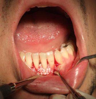
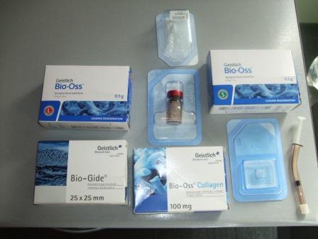
However, when the bone defects are higher, sometimes to the extent that the teeth are broken, a surgical intervention is performed, where bone defects are cleared and the diseased tissue is removed. In order to facilitate the creation of new bones, various bone substitutes are used. artificial bones (Bio-Oss “Geistlich”), membranes (Bio-Gide “Geistlich”) and growth factors, which enable the restoration of broken tissue, and the consolidation of broken teeth.
How successful is the treatment of periodontal disease?
In the treatment of periodontitis we achieve great success, and due to the cooperation with patients, satisfaction is most often mutual. Particularly satisfied are patients who came to late stages of the disease when they were proposed to extract and put in total dentures due to the dental implant. However, after therapy, they retain their own, fixed teeth, which perform both functional and aesthetic role.
Preventive measures to be taken in order not to develop periodontal disease
Prevention is the best way to prevent the onset of the disease, so we pay a lot of attention to it. Preventive measures to be taken in the fight against periodontal disease include:
- Patient talk and training in order to properly maintain hygiene of the mouth and teeth (link to the wider text). This implies tooth washing in the correct technique, after each meal, and at least 3 times a day for 3 to 5 minutes, using brushes and toothpaste as well as dental hygiene aids, ie, interdental end and interdental brushes.
Proper nutrition (link to more extensive text): Consuming fresh fruits, vegetables and abrasive, solid foods. - Regular controls (every 6 months or more) for removing dental calculus, which is normally created, as well as soft teeth from the teeth, and the condition of the gingiva and teeth is controlled.
- It is very important that all the carotid teeth cure. All inadequate prosthetic restraints (crowns, bridges) and sealants, which mechanically irritate the gums, should be removed and replaced with more adequate ones.
If parodontopathy is already present, a therapy that depends on the stage of disease development is applied.
We explain to patients that every, and the smallest symptom you notice on the right, should not be ignored, especially bleeding from the right. For these reasons, we advise them to report to us immediately, as well as to come to regular checks.
CASES GALLERY
AESTHETIC PERIODONTICS – GINGIVECTOMY
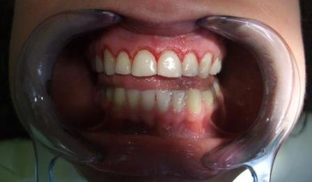
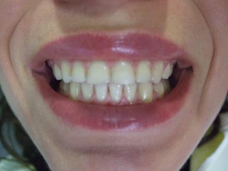
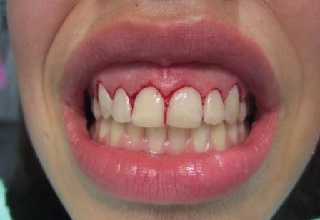
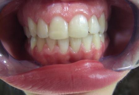
1.Female patient, 27 years old, gingivectomy was made.
Picture 1: Right after the gingivectomy
Picture 2: 2 weeks after the gingivectomy
Picture 3: 6 months after the gingivectomy
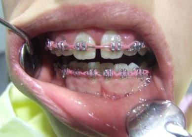
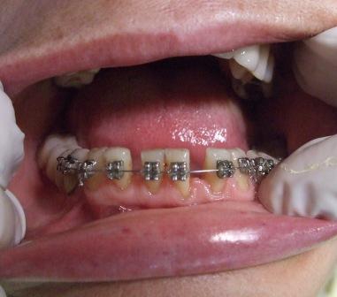
2.Female patient, 16 years old, gingivectomy was made after fixed braces was removed.
Picture 1: After fixed braces was removed
Picture 2: Right after the gingivectomy
Picture 3: 3 weeks after the gingivectomy
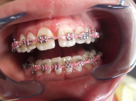
3.Female patient, 13 years old, gingivectomy was made during fixed braces therapy.
Picture 1: Before
Picture 2: After
GINGIVECTOMY
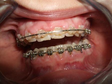
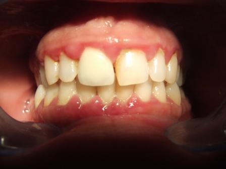
Pacijentkinja, 13 godina, hiperplazija desni.
Picture 1: Before – 2004, terminal stadium of periodontitis
Picture 2: Periodontal therapy, installation of artificial bone and orthodontic treatment were made.
Picture 3: 2010, the patient has all her teeth. Stable condition.
TREATMENT OF TERMINAL PERIODONTITIS WITH TEETH MIGRATION
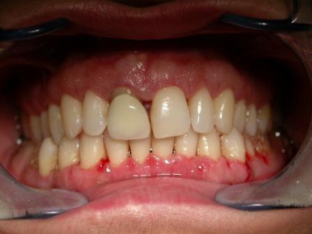
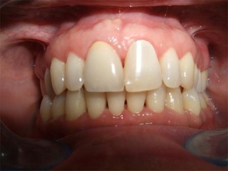
Female patient, in 2004 came with terminal stadium of periodontitis.
Picture 2: After periodontal therapy
Picture 3: After crown replacement
PERIODONTICS – ULCERONECROSIS CHANGES ON THE GUMS WITH TEETH MIGRATION
Female patient, 27 years old.
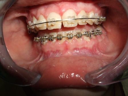
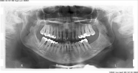
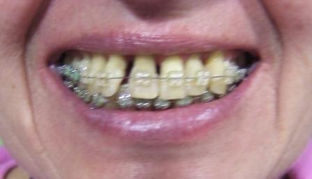
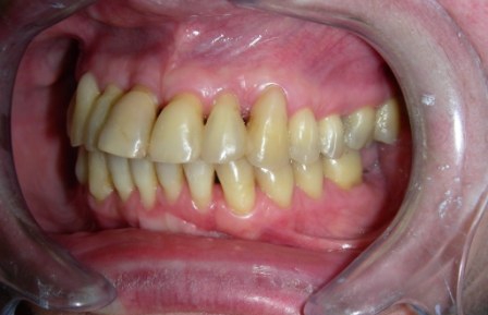
PERIODONTICS – COMBINATION WITH ORTHODONTICS
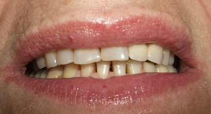
Female patient, 55 years old.
Picture 1: Before
Picture 2: After periodontal therapy fixed braces are placed
Picture 3: After the therapy
PERIODONTICS – INSTALLATION OF ARTIFICIAL BONE
1. Guided tissue regeneration with the application of collagen membrane.
Picture 1: Application of collagen membrane
Picture 2: Collagen membrane in place
2. Installation of artificial bone.
Picture 1: Application of bone transplant
Picture 2: Materials for regenerative therapy
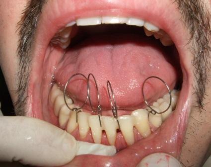
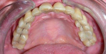
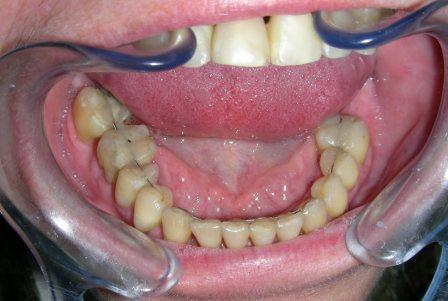
SPLINT – FIXING TEETH IN TERMINAL PERIODONTITIS
Male patient, 30 years old, placing fiberglass splint.
Picture 1: Placing fiberglass splint
Picture 2: DENTAFLEX SPLINT-upper jaw.
Picture 3: DENTAFLEX SPLINT-lower jaw.
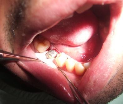
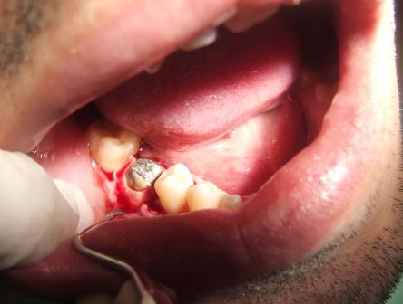
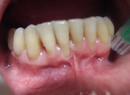
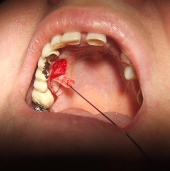
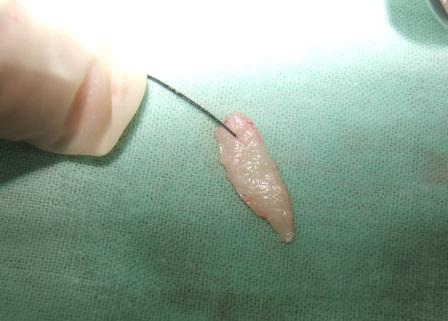
PERIODONTICS- MUCOGINGIVAL SURGER
Picture 1: Narrow and receding gums.
Picture 2: Taking the transplant from the palate.
Picture 3: Transplant from the palate.
Picture 4: A month after the intervention. Further gums receding is prevented.


