What is inside the tooth?
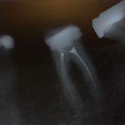
Inside the tooth, under the hard tissue of the lungs and dentin, there is a soft tissue called the pulp or “nerve” of the teeth. It contains except nerves, blood vessels and various types of cells and fibers. The tooth pulp (the “nerve” of the teeth) is surrounded by the hard tissue of the root of the teeth. The pulp spreads up, toward the crown of the tooth, and down – towards the top of the root, where it is in contact with the soft tissue around the root of the teeth. It is very important for the growth and development of teeth. However, if due to infection or other causes, the tooth pulp is removed (removed from the root canal), it can survive without the pulp, as it continues to be fed through the soft tissue that surrounds its root.
Causes of pulmonary disease (“nerve”)
Causes of inflammation or infections of the pulp (“nerve”) are different: deep caries lesion, repeated treatments on the tooth, broken teeth. Dental injuries can also damage the pulp, although no visible signs of its previous damage should be visible on the tooth itself. If inflammation or infection is left untreated, they can cause pain leading to an abscess. However, sometimes the pulp can be irreversibly ill, without any symptoms of pain or sensitivity.
What is endodontic treatment or treatment of the root canal?
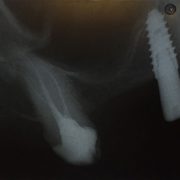
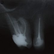
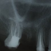
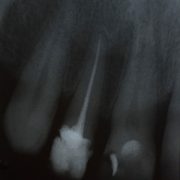
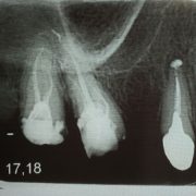
Endodontic treatment or treatment of the root canal channel is necessary when the pulp is inflamed or infected. Endodontic treatment consists of removal of inflamed or infected pulp (“nerve”), and careful cleaning and shaping of the inner part of the root where the pulp was located. After complete mechanical and chemical treatment, the root canal is filled with appropriate pastes and gutta-percha wands.
How is the treatment of the root canal in our clinic conducted?
In our clinic mechanical processing of the root canal is done by special machine tools made of titanium so-called. NiTi Protaper. These instruments allow almost every tooth to be healed. This is especially important in the teeth of the lateral region, where we have two, three or four roots and the same number of channels, which in most cases are curved or inaccessible if they are done with ordinary extensors. The length of the channel itself is determined using the instrument – Apex locator, retroalveolar image, or CD 3D image of a particular tooth. The final filling of the canal is done by cold condensation of non-corrosive paste and gutta-percha.
Each phase of work is explained to patients, so that excellent communication and willingness to cooperate are achieved, which greatly contributes to our and their success in the preservation of teeth. Due to the use of modern and modern devices, as well as appropriate anesthetics, many patients have confirmed that they felt comfortable throughout the entire procedure.
After the treatment of the root canal treatment, restoration, replacement of the crown of the tooth, and restoration of the same to the full function, so that it can continue its role in the mouth, as well as every other tooth.


